Case History
INTRAPAPILLARY INJECTION OF HARVESTED DNA INTO OPTIC NERVE OF BLIND PATIENT
All through medicine we are taught not to hit any nerve especially the optic nerve, however here for the first time we are using an approach we term Intra P apillary injection of DNA right into the optic nerve of blind patients. After completing an eye surgery called vitrectomy where the toxins and bands from the inner depths of the eye are removed using 25 Gauge instruments, the DNA injection is given loaded on a 10 micro milliliter syringe and very thin 26 gauge to 33 gauge needles. The response is immediate where the patient is once again able to see light, since the patients own DNA bioengineered and harvested for this purpose is used.
RETROBULBAR INJECTION INTO OPTIC NERVE OF HARVESTED DNA INTO BLIND PATIENT
Where no surgery is to be conducted the optic nerve can still be approached through the periphery called the retrobulbar space guided by surgeons experience in getting the injection into the optic nerve or its sheaths or coverings. These kind of injections are repeated for better results the need to go intraocular thus being minimized, also this route is used in patients with low vision from hand movements they improve to counting fingers.
PRE AND POST PICTURES OF PATIENT WITH PERCEPTION OF LIGHT NEGATIVE WITH DNA INJECTIONS TURNS PERCEPTION OF LIGHT POSITIVE
Patient with Bilateral Cataract, Uveitis, Glaucoma improved within 10 days of the course of Mancells from no perception of light to perception of light. The patient could actually tell from which direction the light is coming wherein previously he had no perception of light itself.
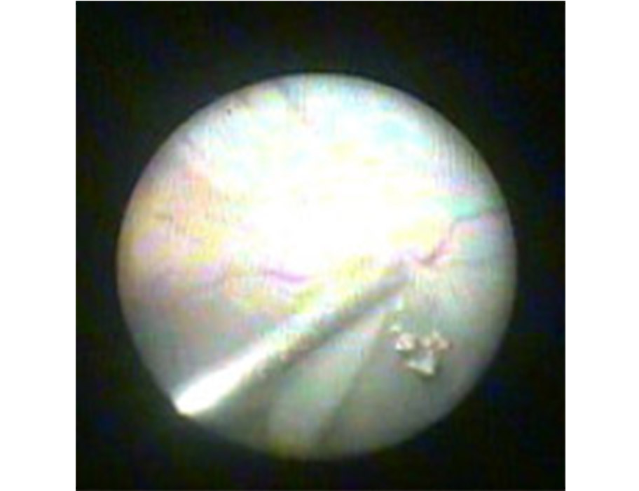
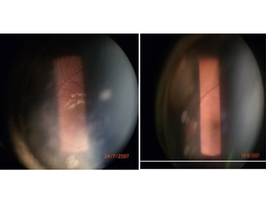
PRE AND POST OF MACULAR DEGENERATION IN YOUNG PATIENT AFTER DNA INJECTIONS
Young man presented with seeing wavy patterns and blind areas within his visual field, on examination it showed the optic nerve and macular to be degenerated. This can be seen as the white patches seen on the pre treatment retinal photograph. After undergoing a course of 10 days with Manacells the retinal improvement could be clearly photographed showing reduction of the white patches and healthy pink glow of the retina, the symptoms of the patient reduced with increased visual field and final vision.
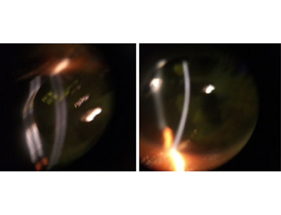
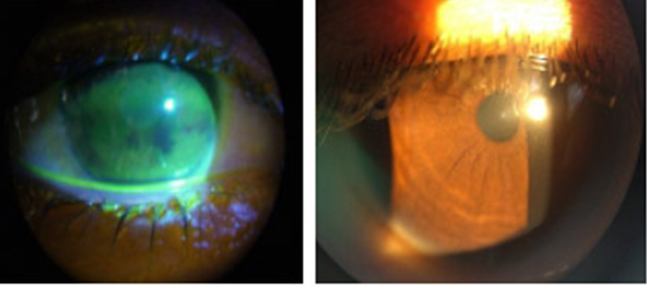
PRE AND POST OF PATIENT WITH KERATOCONUS AFTER MANACELLS THERAPY
Young person presented with blindness and vision of hand movements due to Keratoconous a blinding degeneration of the cornea making it very thin and giving it a cone like shape. This can be seen with the way the light from the slit lamp biomicroscope makes with the cornea or the front window of the eye. The cornea should ideally be over 500 microns (half millimeter) however in the pre treatment photograph the cornea was 378 microns. After the prescribed regime for 10 days of Manacells therapy the corneal thickness improved to 412 microns and the patients vision improved to three lines on the vision chart (6/24). Thus corneal surgery could be avoided.
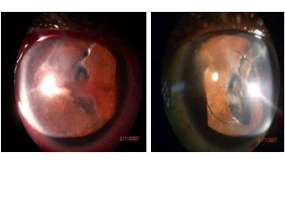
PRE AND POST PHOTOGRAPHS OF DRY EYE PATIENT
Patient with dry eyes showing stainging with fluorescence showing the gaps in the tear film and with 10 day regime of Manacells therapy the patients tear film becomes complete due since it aids and increases the all the three layers of tear film of essential the mucus generated from the goblet cells of the conjunctiva or mucus membrane of the eye and this brings health back to the cornea and the eye, making it comfortable for the patient and increases the patients vision.